Why Your Knees Hurt When You Squat (And How to Fix It)
⚠️ Disclaimer: The information in this article is for general educational purposes only and does not constitute medical, nutritional, or professional fitness advice. Individual results may vary. Always consult a qualified healthcare professional or certified fitness trainer before starting any new exercise program, changing your diet, or making decisions about injury treatment or recovery. If you experience pain, discomfort, or any unusual symptoms during exercise, stop immediately and seek professional guidance.

The Anatomy of the Knee During a Squat
Understanding the specific mechanical demands the squat places on the knee joint — and the structures that experience the greatest stress during those demands — provides the foundation for understanding why and where pain develops.
Understanding what was actually happening inside my knee during a squat made the fix feel logical rather than arbitrary — anatomy context matters.
Knee Joint Structures Involved in Squatting
The knee joint is not a simple hinge — it is a complex structure involving three bones (femur, tibia, patella), four primary ligaments (ACL, PCL, MCL, LCL), the medial and lateral menisci, multiple bursae, and an extensive musculotendinous system that spans the joint from both above and below. During a squat, each of these structures experiences specific mechanical loads that vary with squatting depth, technique, load, and the mobility and strength characteristics of the individual performing the squat. The patellofemoral joint — the articulation between the patella (kneecap) and the femoral trochlea (the groove at the front of the femur where the patella tracks) — experiences compressive forces that increase dramatically with squatting depth: at 0 degrees of knee flexion (standing), patellofemoral joint reaction force is approximately 0.5 times body weight; at 90 degrees of flexion (parallel squat), it reaches approximately 7 times body weight; and at full depth (below parallel), it can exceed 8 times body weight in loaded squats. These compressive forces are not inherently injurious — the patellofemoral joint is designed to handle them — but they become problematic when the patella tracks laterally rather than centrally in the femoral groove, when the cartilage surfaces have developed irregular wear patterns, or when the compressive loading has increased faster than the articular cartilage’s adaptation rate can manage.
The infrapatellar fat pad — a highly innervated fat deposit located immediately below the patella and between the patellar tendon and the joint capsule — is one of the most pain-sensitive structures in the knee and is frequently impinged during squatting movements that compress the anterior knee structures. Fat pad impingement produces a characteristic sharp pain at the bottom of the kneecap that is often confused with patellar tendinopathy (pain in the patellar tendon below the kneecap) but has a distinct location (more central and deeper than tendon pain) and distinct aggravating mechanics (compression rather than tension). The distinction matters for management: patellar tendon pain is primarily managed through eccentric loading and progressive tendon strengthening; fat pad impingement is managed through reducing the terminal extension loading that compresses the fat pad and addressing the quadriceps activation patterns that alter the fat pad’s mechanical environment. Squatting technique that avoids extreme knee hyperextension at the top of the movement — and that maintains a slight knee soft-lock rather than fully locking out — reduces fat pad impingement risk in individuals with established fat pad sensitivity.
Patellofemoral Mechanics During Squatting
Patellofemoral tracking — the path the patella takes as the knee flexes and extends during a squat — is one of the most important biomechanical variables in squatting knee pain development, because lateral maltracking dramatically increases the pressure on the lateral facet of the patella and reduces it on the medial facet, producing the uneven cartilage wear and increased lateral retinaculum tension that patellofemoral pain syndrome involves. Normal patellar tracking follows the central groove of the femoral trochlea through the full range of knee flexion and extension, guided by the balance between the medial and lateral soft tissue forces that pull the patella from each side. Lateral maltracking — the most common tracking pathology — occurs when lateral forces (tight iliotibial band, tight lateral retinaculum, weak vastus medialis oblique) exceed medial forces (vastus medialis oblique contraction, medial retinaculum tension), producing the laterally displaced or tilted patella that compresses the lateral patellar facet and places abnormal stress on the lateral retinaculum. The Q-angle — the angle between the line of pull of the quadriceps and the line of the patellar tendon — provides a rough anatomical predictor of patellar tracking tendency: higher Q-angles (more common in women due to wider pelvic width relative to knee spacing) are associated with greater lateral tracking tendency and higher patellofemoral pain syndrome prevalence, though Q-angle alone is not a sufficient explanation for individual knee pain presentations.
Forces on the Posterior Knee Structures
While anterior knee pain from patellofemoral dysfunction is the most common squatting pain pattern, posterior and medial knee structures also experience significant loads during squatting that produce their own pain presentations in specific circumstances. The posterior cruciate ligament (PCL) experiences peak loading during deep squatting when the posterior tibial translation that the quadriceps contraction produces must be resisted — a loading pattern that is problematic in individuals with PCL laxity or previous PCL injury where the ligament’s mechanical contribution is reduced. The medial collateral ligament (MCL) and the medial meniscus experience increased loading when knee valgus (inward collapse) occurs during squatting — the medial structures are placed under tensile stress as the knee collapses medially, producing the medial knee pain that valgus-pattern squatting generates. The popliteal tendon and posterior capsule can be stressed by the extreme range of motion that full-depth squatting demands in individuals with posterior chain tightness, producing the posterior knee pain that deep-squatting beginners sometimes report. Understanding which structures are producing the specific pain pattern — anterior, medial, lateral, or posterior — is the critical diagnostic step that guides the appropriate intervention, because the same “knee pain during squatting” complaint can reflect entirely different anatomical problems requiring different solutions.
The Role of Hip and Ankle Mechanics
The knee does not function in isolation during a squat — it is mechanically linked to the hip above and the ankle below through the kinematic chain that transmits force from the floor through the entire lower extremity. Hip mechanics profoundly affect knee loading: femoral internal rotation (produced by hip weakness or mobility restriction) creates the knee valgus pattern that places abnormal stress on the medial knee structures; hip extension weakness forces the quadriceps to dominate the squat pattern, increasing patellofemoral loading beyond the levels that hip extensor contribution would share; and hip external rotation restriction limits the foot external rotation and knee-outward positioning that reduces patellofemoral stress in the squat pattern. Ankle dorsiflexion restriction — limited ability to bring the shin forward over the foot during the squat descent — is one of the most prevalent and most overlooked contributors to squatting knee pain, because restricted dorsiflexion forces one of three compensatory patterns: excessive heel rise (shifting weight to the forefoot and dramatically increasing patellofemoral loading), excessive trunk lean (maintaining balance by increasing forward lean, which shifts demand away from the knees but creates compensatory lumbar stress), or excessive knee travel beyond the toes (which the knee-tracking-over-toes technique instruction correctly identifies as problematic but whose actual cause — ankle restriction — the instruction does not address). Addressing ankle dorsiflexion restriction directly, rather than simply cueing the knee not to travel forward, is one of the highest-leverage squatting knee pain interventions available.
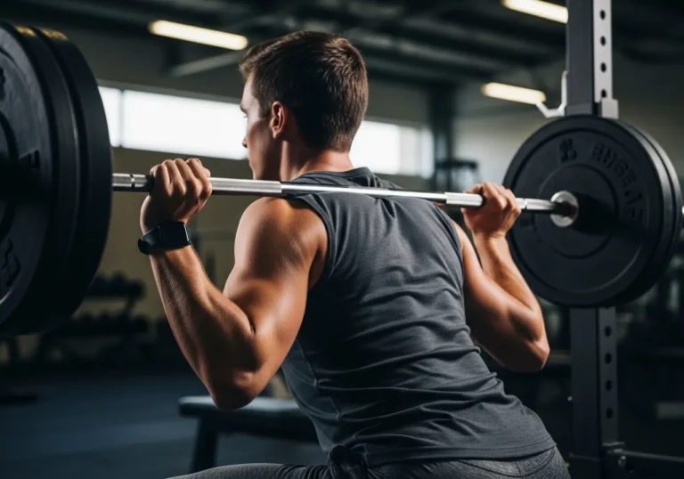
Load Distribution: How Squatting Variables Affect Knee Stress
The specific mechanics of the squat — bar position, stance width, foot rotation, squatting depth, and torso angle — all significantly affect how compressive and shear forces are distributed across the knee joint structures, and understanding these relationships enables technique modifications that reduce knee loading in pain-provoking patterns. High bar squat (barbell positioned on the upper trapezius) produces greater knee flexion at equivalent depth compared to low bar squat (barbell positioned on the posterior deltoid), generating higher patellofemoral compressive forces and more anterior knee dominance. Wider stance squatting reduces the knee flexion required at equivalent depth by increasing hip contribution to the movement, reducing patellofemoral compressive loading while increasing hip joint loading — a trade-off that is favorable for individuals with patellofemoral pain and unfavorable for those with hip pathology. Greater foot external rotation (toes pointed further outward) facilitates hip external rotation and reduces the femoral internal rotation that contributes to valgus collapse, while also requiring greater hip mobility to maintain the knee-over-toe alignment that the externally rotated position demands. These relationships provide specific, actionable technique modification options for individuals with squatting knee pain — making the biomechanical understanding that this section provides the essential foundation for the technique correction strategies that section three describes.
The relationship between training load and knee tissue adaptation — specifically the differential adaptation rates of muscle versus connective tissue — explains why squatting knee pain so frequently emerges during periods of rapid load increase that feel manageable from a muscular strength perspective. Muscle tissue adapts to increased loading within 2 to 4 weeks through the well-known processes of hypertrophy and neural adaptation; cartilage, tendons, and ligaments adapt 8 to 12 weeks or more after equivalent loading increases, because their slower cellular turnover and reduced vascularization limits the rate at which structural remodeling can occur. This mismatch creates a vulnerability window — typically 4 to 8 weeks after a loading increase — during which the muscles feel capable of the load but the passive knee structures have not yet adapted to the mechanical demands the stronger muscles are now capable of imposing. Recognizing this adaptation lag explains why the most important single variable in preventing squatting knee pain is load progression rate rather than any specific technique element — and why the 10 percent weekly load increase guideline that injury prevention research supports is directly applicable to squatting programs, regardless of how much muscular strength has developed beyond the rate that connective tissue adaptation can match.
| Knee Structure | Primary Squat Load | Pain Pattern | Common Cause |
|---|---|---|---|
| Patellofemoral joint | Compressive (increases with depth) | Anterior, around/behind kneecap | Lateral maltracking, rapid load increase |
| Patellar tendon | Tensile (quadriceps pull) | Below kneecap | Overuse, rapid volume increase |
| Medial structures (MCL, meniscus) | Tensile during valgus | Inner knee | Knee valgus collapse |
| IT band / lateral retinaculum | Tensile + compressive | Outer knee | Lateral maltracking, IT band tightness |
| Infrapatellar fat pad | Compressive (terminal extension) | Deep to patellar tendon | Hyperextension, anterior knee compression |
Research published in the Journal of Orthopaedic and Sports Physical Therapy found that the majority of knee pain during squatting is biomechanical rather than structural — caused by correctable technique errors including excessive forward lean, knee valgus collapse, and inadequate ankle dorsiflexion — and that form corrections alone resolve pain in approximately 70 percent of cases without requiring training cessation.

The Most Common Causes of Knee Pain in Squats
Most squatting knee pain falls into a small number of identifiable categories, each with specific causes and specific interventions that address the actual problem rather than simply managing symptoms.
My knee pain turned out to be almost entirely from caving inward at the bottom — a cue I’d been ignoring for months that took two weeks of focused practice to fix.
Patellofemoral Pain Syndrome (PFPS)
Patellofemoral pain syndrome — commonly called “runner’s knee” though equally prevalent in squatters — is the most common cause of anterior knee pain in gym-active adults, accounting for approximately 25 percent of all knee injuries presenting in sports medicine clinics. PFPS is characterized by diffuse anterior knee pain that is worse with squatting, stairs, prolonged sitting with knees bent (the “theater sign”), and activities that load the knee in flexion under compressive force. The pain is typically described as aching or burning around or behind the kneecap, is often bilateral though usually more severe on one side, and is frequently accompanied by a grinding or grating sensation during knee flexion that reflects the altered patellar cartilage surface that chronic PFPS produces over time. The primary causes of PFPS in recreational squatters: rapid increases in squatting volume or load that exceed the patellofemoral joint’s adaptation rate (the most common single cause); lateral patellar maltracking from the muscle imbalances described in section 1-2; quadriceps muscle tightness that increases patellofemoral compressive force throughout the range of motion; hip abductor weakness that allows femoral internal rotation and knee valgus that increases lateral patellar loading; and foot pronation that transmits tibial internal rotation up the kinematic chain to produce femoral internal rotation and lateral patellar stress. The good news for PFPS sufferers: research consistently shows that appropriately directed rehabilitation — specifically addressing the contributing biomechanical factors through the technique corrections, mobility work, and strengthening described in sections three through five — produces complete pain resolution in the majority of PFPS cases within 6 to 12 weeks.
Patellar Tendinopathy
Patellar tendinopathy — sometimes called “jumper’s knee” — is pain in the patellar tendon (the thick tendon connecting the patella to the tibial tuberosity below the knee) resulting from chronic overuse that exceeds the tendon’s adaptive capacity. Unlike PFPS which involves the patellofemoral joint itself, patellar tendinopathy involves the tendon tissue — and the distinction is important because the management approaches are different. Patellar tendinopathy presents with pain that is specifically located at the inferior pole of the patella (the bottom tip of the kneecap) or along the patellar tendon body, is typically worst at the beginning of activity (warming up as activity continues), and worsens with high-load jumping, landing, and deep squatting activities that create high tensile loads in the patellar tendon. The primary cause is repetitive high-load activities that create microtrauma in the tendon faster than the tendon can repair — rapid increases in squatting volume, jump training, or running volume are the typical precipitating events. The management of patellar tendinopathy is fundamentally different from PFPS management: whereas PFPS responds well to unloaded stretching and hip strengthening, patellar tendinopathy requires progressive tendon loading through heavy slow resistance training (specifically isometric and heavy isotonic loading that stimulates tendon collagen remodeling without exceeding the tendon’s current capacity) and is typically worsened by stretching and passive treatments that do not provide the mechanical loading the tendon requires for structural repair.
Knee Valgus: The Most Correctable Cause
Knee valgus — the inward collapse of the knee during squatting that produces the classic “knees caving in” appearance — is simultaneously the most visible and the most correctable cause of squatting knee pain, and addressing it through the technique corrections and strengthening described in sections three and five resolves the medial knee stress that valgus produces in the majority of cases without requiring medical intervention. Knee valgus during squatting results from a combination of contributing factors: hip abductor weakness (the gluteus medius and minimus cannot maintain hip abduction resistance against the adduction forces that valgus collapse represents, allowing the femur to internally rotate and adduct under load); limited hip external rotation mobility (insufficient range of motion for the knee-out position that valgus prevention requires at depth); pronated foot mechanics that transmit tibial internal rotation upward; and insufficient lateral hip engagement cuing during the squat that the individual has not been taught to maintain under load. Each of these contributing factors is addressable: hip abductor strengthening through clamshells, side-lying abductions, and banded squats; hip external rotation mobility through the piriformis and hip rotator stretches described in the lower back pain guide; foot mechanics assessment and correction; and deliberate “push knees out” cuing during squatting practice that builds the motor pattern before load is progressively reintroduced.
Iliotibial Band Syndrome (ITBS) in Squatters
While iliotibial band syndrome is more commonly associated with running, it produces lateral knee pain in squatters through a mechanism that differs somewhat from the running presentation. The IT band — a thick fibrous band running from the hip to the lateral tibia — creates the lateral knee pain of ITBS when it becomes excessively taut and compresses the lateral knee structures during repetitive flexion-extension cycles. In squatters, ITBS-related lateral knee pain is most prominent at the bottom of the squat and during the ascent from depth — the range of motion where the IT band’s compressive load on the lateral femoral condyle is highest. Contributing factors specific to squatters: wide-stance squatting that increases lateral hip tension and IT band load; hip flexor tightness that tilts the pelvis anteriorly and increases IT band tension through its proximal attachment; and foam rolling the IT band itself (which is a tendinous structure, not a muscle, and does not respond to compression the way muscle tissue does — the more effective target is the tensor fascia latae at the hip and the gluteus maximus at the iliac crest, which modulate IT band tension through their proximal attachments).
Pes Anserine Bursitis and Medial Knee Pain
Pes anserine bursitis — inflammation of the bursa located at the medial proximal tibia where the pes anserine tendons (sartorius, gracilis, semitendinosus) attach — produces medial knee pain that is sometimes confused with medial collateral ligament pain or medial meniscus pathology. The characteristic location is approximately 2 to 3 centimeters below the joint line on the medial tibia — distinguishable from MCL pain (which is at the joint line) and from meniscal pain (which is also at the joint line but has additional mechanical symptoms like locking or giving way). Pes anserine bursitis is particularly common in individuals who perform high-volume squatting with significant knee valgus, because the valgus pattern increases the tensile load on the medial knee structures including the pes anserine tendons. Management includes reducing the valgus component of squatting technique, temporarily reducing squatting volume and depth to allow bursal inflammation to resolve, ice application to the specific bursal location, and addressing the hamstring and hip adductor tightness that increases pes anserine tendon tension at the attachment site.
Overuse: The Volume and Load Progression Cause
Overuse — training load increases that exceed the adaptive capacity of the knee’s various structures — is the most common underlying cause of squatting knee pain across all the specific presentations described above. The knee structures most vulnerable to overuse-related damage: the articular cartilage (which has minimal regenerative capacity and accumulates damage from loading that exceeds its viscoelastic properties); the patellar tendon (which adapts to loading more slowly than the quadriceps muscles it connects, creating the adaptation mismatch that tendinopathy exploits); and the subchondral bone beneath the articular cartilage (which responds to overuse with microfractures that produce the bone marrow edema that MRI reveals in overloaded joints). The primary overuse scenarios in recreational squatters: adding more than 10 percent weekly load to squatting programs; returning to previous loading levels after a break without a graduated re-entry; performing high-frequency squatting (daily or near-daily) without adequate recovery between sessions; and combining high-volume squatting with other knee-dominant activities (running, stair climbing, cycling) without accounting for the cumulative knee loading across all activities. The correction is straightforward — temporary load reduction to below the pain threshold, followed by graduated re-progression at a rate that allows adaptation — but requires the patience that most recreational athletes underestimate when they first implement it, as the overuse injuries that months of excessive loading have produced require weeks to months of appropriate loading to resolve.
Chondromalacia patellae — softening and degradation of the articular cartilage on the posterior surface of the patella — is a structural finding on MRI or arthroscopy that is sometimes identified as the “cause” of squatting knee pain but whose relationship to symptoms is more complex than the diagnosis implies. Research consistently shows that chondromalacia patellae is highly prevalent in asymptomatic individuals — up to 30 percent of people without knee pain have MRI evidence of patellar cartilage changes — while symptomatic individuals frequently have normal patellar cartilage on imaging. This disconnect between structural findings and symptoms reflects the well-established principle that pain is not simply a signal of tissue damage — it is a complex neuroscientific phenomenon that involves tissue loading, neural sensitization, psychological factors, and individual pain processing characteristics that structural imaging cannot capture. For recreational squatters told they have chondromalacia patellae and advised to stop squatting, the more evidence-based approach is to address the biomechanical contributors to their specific knee pain pattern through the techniques described in this guide, rather than allowing a structural label to produce permanent movement avoidance that the research on exercise and cartilage health does not support.
| Cause | Pain Location | Key Identifier | Primary Fix |
|---|---|---|---|
| PFPS | Around/behind kneecap | Worse after prolonged sitting | Hip strengthening, load management |
| Patellar tendinopathy | Below kneecap (inferior pole) | Worse at start of activity | Heavy slow resistance loading |
| Knee valgus | Medial knee | Visible inward knee collapse | Glute strengthening + technique |
| ITBS | Lateral knee | Worse at specific squat depth | TFL/glute rolling, hip strengthening |
| Overuse | Variable | Onset after load increase | Reduce load, graduated re-progression |
Research published in the Physical Therapy journal found that targeted hip strengthening — particularly the glute medius and external rotators — reduces knee pain during squatting in 70 to 80 percent of cases within 4 to 6 weeks, without any modification to the squat itself, confirming that knee pain during squats is frequently a hip strength issue rather than a knee problem.

How to Fix Your Squat Form to Eliminate Knee Pain
Form correction is the highest-leverage intervention for the majority of squatting knee pain cases — because most knee pain during squatting reflects technique errors that change how load is distributed across the knee structures rather than structural damage that requires medical treatment.
Recording myself squatting from the front was uncomfortable to watch but immediately showed me three form issues I couldn’t feel from the inside.
The Knee-Out Cue: The Single Most Impactful Correction
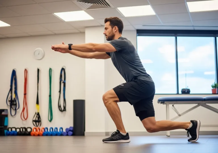
The “push your knees out” cue — actively driving the knees outward in the direction of the little toes throughout the squat descent and ascent — is the single most immediately impactful technique intervention for squatting knee pain, reducing both the knee valgus that stresses medial structures and the lateral patellar maltracking that patellofemoral pain involves. The mechanism: deliberate knee external rotation engagement activates the hip external rotators (primarily gluteus maximus and deep hip rotator group) and the hip abductors (gluteus medius and minimus) that resist the internal rotation and adduction forces that the squat’s loading pattern creates. The cue is most effectively learned through progressive stages: first, perform bodyweight squats with a resistance band around the thighs (just above the knees) that provides resistance to abduction and requires active resistance from the hip abductors to prevent valgus collapse — the band teaches the motor pattern of active knee-out engagement that the unloaded squat should then replicate; second, practice paused squats (stopping at the bottom position for 2 to 3 seconds) where the knee-out engagement can be deliberately checked without the momentum that the full-speed squat involves; third, film the squat from the front and compare the knee position to the foot’s external rotation angle — the knees should track in the direction of the foot, neither caving medially nor pushing excessively laterally beyond the little toe. This correction alone resolves squatting knee pain in a significant proportion of the cases where valgus pattern is the primary contributing factor — making it the starting point for any squatting knee pain correction protocol before the more specific interventions described below are considered.
Addressing Heel Rise: The Ankle Fix
Heel rise during squatting — the compensatory elevation of the heel when ankle dorsiflexion is insufficient to maintain flat-footed contact through the full squat depth — is one of the most common but most overlooked contributions to squatting knee pain that technique correction can address. When the heel rises, the weight distribution shifts to the forefoot, the center of mass moves forward, and the quadriceps must compensate with dramatically increased force to maintain the squatting pattern — increasing patellofemoral compressive loading by 20 to 40 percent compared to flat-footed squatting at equivalent depth. Identifying heel rise: place a small plate (2.5 to 5 kilograms) under the heels during squatting — if this eliminates or significantly reduces knee pain, ankle dorsiflexion restriction is contributing to the pain pattern and direct ankle mobility work is indicated. The immediate modification: elevated heel squatting (using heel elevation or dedicated Olympic weightlifting shoes with built-in heel elevation) allows pain-free squatting while ankle mobility is progressively improved through the specific mobilizations described in section four. The heel elevation is not a permanent solution but a rehabilitation accommodation that allows continued squatting training while the underlying mobility restriction is addressed — typically requiring 4 to 8 weeks of consistent ankle mobility work before flat-footed squatting becomes comfortable.
Stance Width and Foot Position Adjustments
Adjusting stance width and foot rotation provides immediate pain reduction for many squatters by changing the mechanical demands on the knee without addressing the underlying mobility or strength issues — making these adjustments valuable short-term accommodations while the more fundamental corrections described in other sections are developed. Wider stance reduces knee flexion requirements at equivalent depth, decreasing patellofemoral compressive loading — a modification that typically reduces anterior knee pain immediately in patellofemoral pain sufferers. Greater foot external rotation (toes turned further outward, typically to 30 to 45 degrees) facilitates the hip external rotation that valgus prevention requires and reduces the femoral internal rotation that lateral patellar tracking produces — though it requires corresponding hip external rotation mobility that individuals with hip restriction may not have available. The appropriate stance width and foot rotation are not universally prescribed angles but individually determined positions that allow the individual’s hip anatomy (specifically the acetabular depth, femoral neck angle, and hip socket orientation that vary substantially between individuals) to move through full squatting range of motion without impingement, pain, or compensation. Finding the individual stance through stance width experimentation — beginning with shoulder-width stance and feet at 20 to 30 degrees external rotation, then adjusting width and rotation until the most comfortable, compensation-free position is identified — is more appropriate than prescribing the “correct” stance that population-average anatomy would suggest.
Depth Management: Training to Comfortable Range
Squatting depth is one of the most debated variables in squatting technique, and the optimal depth for pain management requires understanding the relationship between depth and patellofemoral loading described in section 1-1. For individuals with significant patellofemoral pain, temporarily limiting squat depth to the range at which patellofemoral compressive force is tolerable — typically the parallel depth (thighs parallel to the floor) or slightly above — and progressively increasing depth as pain tolerance and patellofemoral adaptation improve, produces better long-term outcomes than either extreme (refusing to squat beyond the pain-free range that may be very shallow, or forcing full depth squatting despite significant pain). The depth modification protocol: begin squatting to the first point at which discomfort begins (not pain — discomfort), and continue to that consistent target depth across 2 to 3 weeks; if this depth is consistently comfortable across the weeks, increase depth by approximately 2 to 3 centimeters and maintain this new depth for another 2 to 3 weeks; continuing this graduated depth progression allows the patellofemoral structures to adapt to progressively greater compressive loading in a managed, controlled way that prevents the symptom flares that rapid depth increases produce.
Weight Distribution: Heel-Dominant Squatting
Many recreational squatters — particularly those with a high-bar, upright-torso squatting pattern — naturally distribute weight toward the forefoot during squatting, increasing quadriceps dominance and patellofemoral loading relative to the hip-dominant, heel-loaded squatting pattern that reduces anterior knee stress. Deliberately shifting weight toward the heels during squatting — by thinking “press the floor away through your heels” or “keep your big toe touching the floor but feel your heel pressing down” — engages the posterior chain (hamstrings, glutes) more effectively in the movement and reduces the proportion of the total force requirement that the quadriceps must generate. The practical test: can you wiggle your toes at the bottom of the squat while maintaining balance? If not, weight has shifted too far forward and heel-dominant cuing is warranted. This modification directly reduces patellofemoral compressive force by reducing the quadriceps force requirement and simultaneously increases hip extensor contribution that distributes the total squatting load more evenly across the lower extremity — producing less knee stress at equivalent squatting intensity.
Box Squats and Tempo Squats for Pain Reduction
Box squats — squatting to a box or bench that stops the descent at a controlled depth — provide a pain-managed squatting alternative that allows continued training while knee pain is being addressed through the technique and mobility corrections described in this section. The box prevents the uncontrolled bottom position that often produces the greatest patellofemoral pain, creates a consistent depth target that eliminates the depth variation that pain provocation inconsistency produces, and allows deliberate technique focus at the depth where pain has previously occurred — enabling the specific motor pattern corrections that the unmanaged squat obscures. Tempo squats — performing the descent at a deliberately slow pace (3 to 5 seconds for the eccentric phase) — improve neuromuscular control of the knee valgus and weight distribution variables that rapid-tempo squatting does not allow time to correct, and reduce the dynamic loading rate that contributes to patellofemoral pain in ballistic squatting patterns. Both box squatting and tempo squatting can be incorporated into the rehabilitation period as training-accommodating modifications that allow continued squat pattern training without the loading that full-speed, full-depth squatting imposes on the painful structures — providing the practice volume that technique correction requires while limiting the tissue stress that pain management demands.
The role of the VMO — vastus medialis oblique, the teardrop-shaped muscle at the inner side of the quadriceps just above the medial knee — in patellar tracking and squatting knee pain management is one of the most discussed topics in patellofemoral pain rehabilitation. The VMO is the primary medial patellar stabilizer — its contraction provides the medial force that counterbalances the lateral forces pulling the patella outward during quadriceps contraction — and strengthening it specifically has been a cornerstone of PFPS rehabilitation programs for decades. The most important VMO activation during squatting occurs in the final 30 degrees of knee extension (the terminal extension range), making the top portion of the squat ascent — not the deep flexion — the phase where VMO engagement most directly affects patellar tracking. VMO-specific technique cues during squatting: “squeeze the inner thigh as you stand up” (activates the medial quad in the terminal extension phase); terminal knee extension exercises using a resistance band (standing with band behind the knee, slowly straightening the knee against band resistance — activates the VMO in isolation without patellofemoral compressive loading); and step-ups (specifically the eccentric control of the descent from a step, which requires high VMO activation in the terminal extension range through eccentric contraction). These targeted VMO activation strategies, combined with the hip strengthening described in section five, provide comprehensive patellar tracking correction that pure hip-focused rehabilitation cannot achieve alone.
| Technique Fix | Pain Pattern Addressed | Implementation |
|---|---|---|
| Knee-out cue | Medial knee, PFPS | Banded squats → cued practice → loaded application |
| Heel elevation | Anterior knee, PFPS | Plates under heels while ankle mobility improves |
| Wider stance | Anterior knee, PFPS | Find comfortable stance experimentally |
| Heel-dominant weight distribution | Anterior knee, patellar tendon | “Press through heels” cue, toe-wiggle test |
| Depth reduction | All anterior knee pain | Grad. depth increase every 2–3 weeks |
A study in the Journal of Orthopaedic and Sports Physical Therapy found that ankle dorsiflexion restriction — limited upward ankle mobility — is present in approximately 65 percent of individuals with knee pain during squatting, and that targeted ankle mobility work resolves knee symptoms in a significant proportion of these cases without any direct knee treatment required.

Mobility and Flexibility Work for Healthier Knees
Mobility restrictions are the root cause of many squatting technique problems — and addressing them directly is more effective than indefinitely accommodating them through technique modifications that mask rather than resolve the underlying limitation.
Adding 10 minutes of ankle and hip mobility work before squatting eliminated almost all of my knee discomfort within three weeks — the results surprised me with how fast they came.
Ankle Dorsiflexion Mobilization
Ankle dorsiflexion — the ability to bring the shin forward over the foot with the heel remaining flat — is the mobility restriction most directly linked to squatting knee pain, and improving it produces immediate and lasting improvements in squatting mechanics that reduce knee stress. The standard assessment: stand facing a wall, toes 10 centimeters from the wall, and attempt to touch the knee to the wall while keeping the heel flat. If the knee cannot touch the wall at this distance while the heel remains flat, ankle dorsiflexion restriction is present and is likely contributing to squatting mechanics problems. The most effective ankle dorsiflexion mobilizations: the kneeling ankle rock (kneeling in a lunge position with the front foot flat, drive the front knee forward over the toes while keeping the heel down — 10 to 15 repetitions of this joint mobilization with overpressure from the body weight significantly improves dorsiflexion within a single session, with repeated daily practice producing lasting mobility gains over 4 to 8 weeks); the ankle banded distraction mobilization (using a resistance band anchored at the floor to pull the talus posteriorly while performing the knee-forward movement — addressing the talar positional restriction that limits dorsiflexion range in many individuals who have had ankle sprains); and calf stretching — both gastrocnemius stretch (straight-knee calf stretch) and soleus stretch (bent-knee calf stretch) — which addresses the muscular components of dorsiflexion restriction that the joint mobilizations above do not target.
Hip External Rotation Mobility
Hip external rotation mobility — the ability to rotate the femur outward in the hip socket — is the second most important mobility variable for squatting knee health, because insufficient external rotation limits the knee-out position that valgus prevention requires and forces compensatory internal rotation and valgus that stresses the medial knee structures. Assessment: lie on your back with one knee bent at 90 degrees and the foot raised, then allow the bent knee to fall outward toward the floor (external rotation) — normal range is approximately 45 degrees; if the bent leg cannot reach 45 degrees of outward fall without the pelvis rotating, hip external rotation restriction is present. The most effective hip external rotation mobilizations: the 90/90 hip stretch (sitting on the floor with both legs bent at 90 degrees — one in front, one to the side — and alternating between forward and lateral torso positions that stress the hip into external and internal rotation); the pigeon pose (the classic yoga hip opener that places the hip in combined flexion and external rotation, held for 60 to 90 seconds per side); and the figure-4 piriformis stretch described in the lower back pain guide. Daily practice of these mobilizations for 4 to 8 weeks produces meaningful improvements in hip external rotation that directly improve knee alignment during squatting.
Quadriceps and Hip Flexor Flexibility
Quadriceps tightness increases patellofemoral compressive force by maintaining higher passive tension in the quadriceps-patellar tendon-patella system throughout the range of motion — amplifying the compression that squatting’s active quadriceps contraction produces. Hip flexor tightness (particularly rectus femoris tightness, as it crosses both the hip and the knee) compounds this effect by simultaneously limiting hip extension and increasing the tension in the quadriceps system. The most effective stretches: the standing quadriceps stretch (standing on one leg, pulling the opposite foot toward the buttock while maintaining an upright torso and slight posterior pelvic tilt — 60 seconds per side); the kneeling hip flexor stretch with quadriceps addition (from the kneeling lunge position, reach back with the same-side hand to grasp the rear foot and bring it toward the buttock — simultaneously stretching hip flexor and rectus femoris); and the prone quadriceps stretch (lying face-down, reaching back to grasp the ankle and bring the heel toward the buttock while maintaining neutral lumbar position — providing the most isolated quadriceps stretch available without the hip flexor component that the standing version includes). Each stretch held for 60 seconds per side, performed twice daily during the acute pain management phase, measurably reduces patellofemoral compressive loading within 2 to 4 weeks of consistent practice.
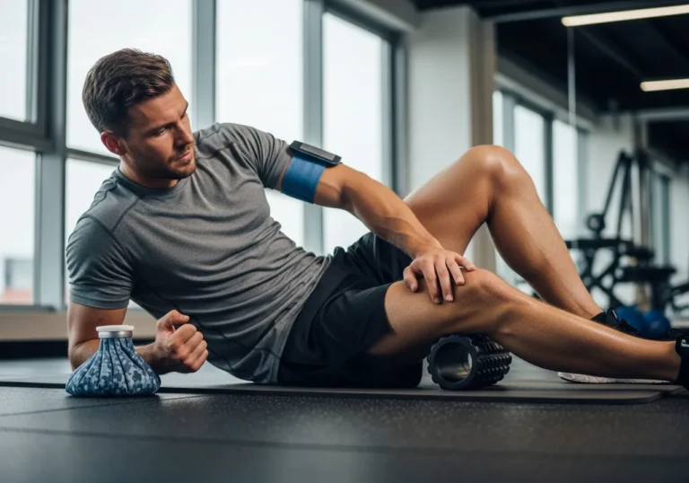
IT Band and TFL Mobility Work
The iliotibial band itself cannot be meaningfully stretched or compressed to therapeutic effect — it is a dense, poorly extensible connective tissue structure whose tension is primarily determined by the muscular attachments at its proximal end (tensor fascia latae and gluteus maximus) and the bony architecture of the lateral knee. Effective IT band tension management therefore targets the tensor fascia latae (TFL) and gluteus maximus rather than the IT band itself. TFL release: foam rolling or lacrosse ball pressure on the upper lateral thigh just below the iliac crest — not the IT band region itself — addresses the TFL muscle belly that modulates IT band tension. Gluteus maximus release: foam rolling the posterior hip with the hip in slight internal rotation (crossed-leg position while rolling) accesses the posterior gluteus maximus fibers that contribute to IT band tension through their fascial connection to the IT band’s proximal attachment. TFL stretching in the lateral lunge position (lunging sideways while keeping the rear leg straight and pushing the hip outward) provides the sustained lengthening that muscle tissue — unlike the IT band itself — can accommodate. These proximal interventions address IT band tension more effectively than the band-rolling approach that most gym-goers employ and that mechanically cannot achieve meaningful IT band lengthening.
Thoracic Spine Mobility and Its Knee Connection
The thoracic spine’s mobility — its ability to extend and rotate freely — has a less obvious but meaningful connection to squatting knee mechanics through the upper body positioning that thoracic mobility enables. Thoracic extension restriction forces a forward-flexed torso position during squatting that shifts the center of mass anteriorly — requiring greater forward knee travel to maintain balance and increasing patellofemoral compressive loading as a compensatory consequence. Improving thoracic extension mobility through the foam roller thoracic extension mobilization (lying back over a foam roller positioned across the thoracic spine, with the roller moving from the lower thoracic to the upper thoracic progressively) and the thread-the-needle rotation stretch described in the lower back pain guide directly improves the upright torso position that reduces anterior knee stress during squatting. Athletes who spend prolonged periods seated tend to develop thoracic extension restriction that compromises squatting mechanics — making thoracic mobility work a surprisingly important component of squatting knee pain management for the desk-working athlete whose rounded upper back has become the default resting posture that carries into their squatting movement pattern.
The relationship between breathing mechanics and knee valgus during squatting is an unexpected but practically important connection that advanced rehabilitation practitioners have documented. Dysfunctional breathing patterns — specifically the pattern of chest-dominant breathing with poor diaphragmatic and pelvic floor coordination — impair the intra-abdominal pressure management that trunk stabilization requires, and reduced trunk stability during squatting forces the lower extremities to compensate with the valgus collapse and foot pronation that increase knee stress. Restoring diaphragmatic breathing mechanics — specifically the ability to generate intra-abdominal pressure through coordinated diaphragm, pelvic floor, and deep abdominal engagement — before squatting provides the trunk stability foundation that prevents the lower extremity compensations that produce valgus. The practical breathing assessment: in a standing position, take a deep breath and observe where the rib cage moves — if only the chest rises and the abdomen remains flat, diaphragmatic function is limited and breathing retraining is indicated. The correction: 360-degree breathing practice — lying on your back, place hands on the lower ribs and attempt to expand them in all directions (front, sides, and back) with each inhalation while the abdomen rises simultaneously — retrains the breathing pattern that trunk stabilization and knee valgus prevention both require.
Foot mechanics — specifically foot pronation (rolling inward) and supination (rolling outward) during squatting — transmit rotational forces up the kinematic chain that directly influence knee valgus and patellofemoral tracking. Excessive pronation during squatting produces tibial internal rotation, which drives femoral internal rotation and knee valgus through the closed-chain kinematic linkage of the lower extremity — making foot position and foot mechanics relevant contributors to squatting knee pain that most knee-focused rehabilitation protocols fail to assess. The foot tripod position — weight distributed across the three contact points of the big toe mound, the little toe mound, and the heel — provides the neutral foot mechanics that minimizes the unwanted tibial rotation from pronation. Actively maintaining the foot tripod during squatting (thinking “press down through all three corners of the foot equally”) provides an immediate, accessible foot mechanics intervention that improves knee alignment in many squatters, particularly those who consistently pronate during loaded movement. For squatters with structural foot pronation that voluntary control cannot adequately address, custom or semi-custom orthotics (insoles that provide medial arch support) can reduce the tibial rotation that flat-footed squatting produces — an intervention that physical therapists and sports podiatrists can assess and prescribe based on individual foot mechanics.
| Mobility Target | Assessment | Key Exercise | Expected Timeline |
|---|---|---|---|
| Ankle dorsiflexion | Knee-to-wall test (<10cm = restricted) | Kneeling ankle rock × 15 reps daily | 4–8 weeks |
| Hip external rotation | Supine external rotation <45° = restricted | 90/90 stretch, pigeon pose 60–90 sec | 6–10 weeks |
| Quadriceps/hip flexors | Prone heel-to-buttock gap | Kneeling hip flexor + quad stretch 60 sec | 4–6 weeks |
| TFL / IT band tension | Lateral knee pain with squat | TFL foam roll + lateral lunge stretch | 2–4 weeks |
| Thoracic extension | Rounded upper back in squat | Foam roller thoracic extension | 4–6 weeks |

Knee-Friendly Squat Variations to Try
During the rehabilitation period — while technique corrections are being practiced and mobility restrictions are being addressed — knee-friendly squat variations allow continued training without the knee stress that the full barbell squat imposes in its uncorrected form.
Box squats saved my training when I was rehabbing knee discomfort — they removed the ambiguity about depth and let me rebuild confidence in the movement.
Goblet Squat: The Best Rehabilitation Squat
The goblet squat — holding a dumbbell or kettlebell at chest height with both hands, then squatting to depth — is the most broadly recommended rehabilitation squat for individuals with squatting knee pain, and for good reason: the anterior weight placement naturally produces a more upright torso, the light load relative to barbell squats reduces total knee compressive force, and the counterbalance effect of the front-held weight makes it easier to sit into depth with heels flat — directly practicing the mechanics that reduce knee stress while minimizing the load that would provoke symptoms. The goblet squat also functions as an excellent teaching tool for the hip-driven squat pattern: “sit between your legs” cuing during the goblet squat naturally produces the hip-dominant movement that reduces quadriceps dominance and anterior knee loading, teaching the pattern that translates to barbell squatting once pain has resolved. Progressions: begin with 5 to 10 kilogram dumbbell goblet squats to comfortable depth; progress to 15 to 20 kilograms as technique solidifies; advance to Bulgarian split squats or front squats as a bridge toward full back squat return.
Box Squat: Controlled Depth, Reduced Tension
The box squat — squatting to a box or bench at the target depth, briefly sitting on the box to eliminate the stretch-shortening cycle, then standing — removes the dynamic bottom position that produces the greatest patellofemoral stress in conventional squatting by eliminating the elastic energy storage and rapid direction change that the bottom of a barbell squat involves. This makes box squatting significantly more comfortable for patellofemoral pain sufferers while maintaining the squat pattern practice that rehabilitation requires. The box height should be set at the depth that allows comfortable, pain-free squatting — typically slightly above parallel — and progressively lowered as pain resolves and mobility improves. An important technique note: the box squat requires the weight to shift backward onto the box at the bottom position (unlike a touch-and-go squat where the box is merely touched), which demands more hip extensor contribution and naturally reduces the forward knee travel that contributes to patellofemoral pain. The brief box sit also provides a checkpoint for technique assessment — at the bottom position, knee alignment, weight distribution, and torso position can be deliberately evaluated and corrected before the concentric phase begins.
Wall Sit: Isometric Loading for Tendon Management
Wall sits — isometric squatting against a wall at a fixed knee angle — are particularly valuable for patellar tendinopathy management, where the emerging evidence strongly supports isometric loading as the most effective acute pain-reducing intervention available. Research published in the British Journal of Sports Medicine showed that a single 45-second wall sit at 70 to 80 percent of maximum tolerated intensity produces immediate reductions in patellar tendon pain lasting up to 45 minutes — a pain-inhibitory effect that allows subsequent training to be performed at higher intensity than it would be possible without the isometric pre-treatment. The mechanism: sustained isometric loading activates cortical inhibitory pathways that reduce the central sensitization contributing to tendon pain, providing a window of reduced pain sensitivity within which rehabilitation exercises and training can be performed more productively. Standard protocol: 5 repetitions of 45-second wall sits at 70 to 80 degrees of knee flexion (deeper = greater patellar tendon loading), with 2-minute rests between repetitions, performed before training sessions to manage patellar tendon pain during the session.
Leg Press: Knee Loading Without Axial Compression
The leg press machine — seated or semi-reclined, pressing a weighted platform away from the body with the feet — provides knee-joint loading that strengthens the quadriceps and posterior chain without the axial spinal loading that barbell squatting imposes, making it a valuable rehabilitation exercise for individuals whose knee pain is worsened by the combined spinal and knee loading of barbell squatting. The leg press allows foot position manipulation (wider stance, greater foot external rotation, higher foot placement) that immediately changes the quadriceps-to-glute contribution ratio and reduces knee-dominant loading — providing the foot position flexibility that fixed-foot squat patterns cannot offer. Caution: very low foot placement on the leg press platform concentrates loading in the quadriceps and dramatically increases patellofemoral compressive force — use mid-to-high foot placement on the platform to shift loading toward the hip extensors that share the compressive knee demand. The leg press is a rehabilitation tool and a training accommodation, not a long-term replacement for the free squat pattern — its primary value is maintaining quadriceps and hip extensor strength while the biomechanical corrections that allow comfortable barbell squatting are developed.
Split Squats and Lunges: Unilateral Training
Split squats (rear foot elevated, Bulgarian split squat) and lunges provide unilateral squat pattern training that allows the more mobility-capable or pain-free leg to be trained while the more restricted or painful leg receives a lower-stress unilateral stimulus. Unilateral training has the additional advantage of revealing bilateral asymmetries — in both mobility and strength — that bilateral squatting masks through the stronger leg’s compensation for the weaker one. The split squat positions the knee in a more controlled trajectory than bilateral squatting because the stable rear foot position limits the compensatory movements that bilateral squats allow, making it an excellent technique development exercise for individuals who struggle to maintain knee alignment in bilateral squatting. For individuals with knee pain that is asymmetric (worse on one side), the Bulgarian split squat allows loading of the less-affected leg at higher intensities while the more-affected leg receives lower-intensity rehabilitation work — maintaining training stimulus on the unaffected side without the bilateral compromise of loading both legs at the intensity appropriate for the injured one.
Landmine squats — performed with a barbell anchored at one end in a landmine attachment or corner, holding the free end with both hands while squatting — provide a unique movement pattern that naturally encourages the hip-dominant, upright-torso mechanics that reduce anterior knee loading while providing enough resistance for genuine strengthening stimulus. The arc-shaped resistance path of the landmine squat guides the torso into a more upright position than conventional barbell squatting allows and makes the knee-out alignment more achievable by the guided movement path. For squatters whose patellofemoral pain is specifically worsened by the forward lean that their high-bar back squat pattern produces, the landmine squat provides a loaded squatting alternative that maintains the upright torso without requiring the deep hip mobility that front squats demand — making it particularly accessible for individuals with both anterior knee pain and limited hip mobility who find the front squat position uncomfortable for additional reasons.
Resistance band squats — performing squatting variations with a loop resistance band around the thighs, just above the knees — serve a dual purpose as both a rehabilitation exercise and a VMO and glute activation tool that directly addresses two of the primary contributors to squatting knee pain simultaneously. The band’s resistance to knee adduction (inward movement) requires active hip abductor and external rotator engagement throughout the squat to prevent the valgus collapse the band is resisting — training the exact motor pattern that healthy squatting knee alignment requires. Research on resistance band squatting and glute activation shows significantly higher gluteus medius and gluteus maximus activation during banded squats compared to identical squats without the band — explaining the band’s effectiveness as both a rehabilitation tool and a warm-up activation exercise before heavier squatting. The band does not need to provide high resistance to produce this activation effect — a light loop band providing approximately 5 to 10 kilograms of resistance to knee adduction is entirely sufficient to elicit the enhanced glute and hip abductor activation the research demonstrates, and progression to heavier band resistance should be gradual and deliberate to allow the hip abductors sufficient time to develop the specific functional strength that the heavier band’s greater resistance demands at full squat depth.
Heel-elevated goblet squats — goblet squats performed with the heels elevated on a pair of 5 to 10 kilogram plates — represent the most appropriate starting point for individuals whose squatting knee pain is accompanied by significant ankle dorsiflexion restriction, combining the goblet squat’s light load and natural torso position with the heel elevation that immediately allows pain-free squatting mechanics while ankle mobility is being developed in parallel. As ankle dorsiflexion improves with the mobility work described in section four, the heel elevation can be progressively reduced — first to 5 kilogram plates, then to 2.5 kilogram plates, then to flat-footed squatting — providing a graduated transition from the elevated-heel accommodation to the flat-footed target mechanics that represents the end goal of ankle mobility rehabilitation. This progressive heel reduction schedule provides objective feedback on ankle mobility progress — specifically, whether the previous heel elevation can be eliminated without knee pain returning — that complements the subjective knee-to-wall assessment described in section four and provides the concrete, session-by-session evidence of improvement that motivates the continued daily ankle mobility practice that the visible, measurable progress in squatting independence makes tangible and rewarding to maintain consistently.
| Variation | Best For | Key Benefit |
|---|---|---|
| Goblet squat | PFPS, general rehab | Natural upright torso, light load, technique teaching |
| Box squat | PFPS, depth management | Controlled bottom position, hip-dominant pattern |
| Wall sit | Patellar tendinopathy | Isometric pain inhibition before training |
| Leg press | All knee pain during loading | No axial load, foot position adjustable |
| Split squat / lunge | Asymmetric pain, technique | Unilateral loading, asymmetry revelation |

When Knee Pain During Squats Is Serious
The majority of squatting knee pain responds to the technique corrections, mobility work, and exercise modifications described in this guide — but a minority of presentations indicate more serious pathology that requires medical evaluation before continuing to load the knee.
I ignored sharp knee pain for three weeks before seeing a physio, who told me I’d made things significantly worse — that mistake cost me two extra months of recovery.
Red Flag Symptoms Requiring Immediate Medical Evaluation
Certain knee symptoms that occur during or after squatting indicate serious structural pathology that should not be managed with the self-directed approach described in this guide. Seek immediate medical evaluation for: significant knee swelling that appears within hours of training (effusion suggests internal derangement — meniscal tear, ligamentous injury, or bone injury — rather than the benign tissue irritation that mild puffiness after training represents); a “pop” or “snap” sensation at the moment of knee injury, particularly if accompanied by immediate instability (the knee feeling like it “gives way”) — the classic mechanism of ACL tear that requires urgent orthopedic evaluation; knee locking — inability to fully extend the knee from a flexed position — which suggests a mechanical block from a displaced meniscal tear or loose body that requires surgical evaluation; knee giving way under load without a specific injury mechanism, suggesting ligamentous laxity or significant meniscal dysfunction that self-directed rehabilitation cannot address; and severe pain that is disproportionate to the apparent injury and does not reduce meaningfully with rest, elevation, and ice within 24 to 48 hours — suggesting pathology beyond the muscle and tendon irritation that those first aid measures typically resolve.
Symptoms Indicating Orthopedic Assessment Is Warranted
Beyond the acute red flags above, several sub-acute presentations warrant orthopedic or sports medicine assessment rather than continued self-management. Pain that has not improved meaningfully after 6 to 8 weeks of systematic implementation of the technique corrections, mobility work, and exercise modifications described in this guide — when the individual has been genuinely consistent and compliant with the program — suggests that either the diagnosis is incorrect (a structural pathology requiring specific management is producing the pain rather than the biomechanical issues this guide addresses) or the contributing factors are more complex than the self-assessment has identified. Pain accompanied by persistent swelling that does not fully resolve between training sessions suggests ongoing structural irritation beyond the typical tendon and joint capsule reaction that settles completely with rest. And any knee pain in an individual with a previous significant knee injury (ACL reconstruction, meniscectomy, previous bone fracture) warrants lower threshold for professional reassessment, as the altered biomechanics and structural changes that previous injury produces create pain patterns that may not respond predictably to the standard interventions this guide describes.
Osteoarthritis and Squatting: The Evidence
A common concern among older recreational athletes is whether squatting accelerates knee osteoarthritis — the cartilage degeneration that produces the chronic knee pain and functional limitation affecting a significant proportion of adults over 50. The evidence on squatting and knee osteoarthritis progression is more nuanced and more positive than the common clinical advice to “avoid squatting with knee arthritis” suggests. Research on exercise and knee osteoarthritis consistently shows that appropriate strength training — including squatting — reduces pain, improves function, and slows cartilage degeneration in people with established knee osteoarthritis, with the muscle strength improvements that training produces providing greater joint protection than the loading reduction that avoiding squatting would create. The key qualifiers: “appropriate” squatting for the osteoarthritic knee means loads and volumes that do not produce significant symptom flares, depths that remain within the pain-free range, and the technique modifications described in this guide that reduce peak joint stress — not the pain-through-it approach that can accelerate degeneration, but not the complete avoidance that allows the muscle atrophy and joint instability that accelerates degeneration through different mechanisms. The most appropriate resource for older adults with diagnosed knee osteoarthritis who wish to continue squatting is a sports medicine physician or physiotherapist with experience in osteoarthritis management who can provide the individualized exercise prescription that the heterogeneity of osteoarthritis presentations requires.
Building Long-Term Knee Resilience
Long-term knee health in the recreational squatter is the product of the consistent application of the principles described across this guide: technique that distributes knee loading appropriately, mobility that supports the movement patterns technique requires, strength that actively stabilizes the joint through the full squatting range, and load management that allows the knee’s various structural components to adapt to progressively greater demands without exceeding their adaptive capacity at any point. The knee that is pain-free, strong, and mobile after 10 or 20 years of regular squatting is not the knee that was lucky — it is the knee whose owner invested consistently in the technique, mobility, and strength practices that durable knee health requires. Building these practices into the regular training routine — the prehabilitation exercises before each session, the mobility work that the warm-up includes, the technique cues that each set incorporates, and the load management that the training program applies — is the investment that separates the decades-long squatter from the recurring-injury squatter who cannot maintain training continuity across the years that genuine fitness development requires.
Return-to-Squatting Protocol After Knee Pain
The return to full barbell squatting after a period of knee pain management follows a structured protocol that prevents the re-provocation of symptoms that premature return to previous loads produces. Phase 1 (weeks 1 to 2): goblet squats and box squats at comfortable depth, body weight to light load, focusing entirely on technique quality — knee alignment, heel contact, weight distribution, torso angle. Phase 2 (weeks 3 to 4): front squats or safety bar squats with moderate load (50 to 60 percent of pre-pain working weight), maintaining full range of motion within pain-free limits. Phase 3 (weeks 5 to 6): back squat return at 60 to 70 percent of pre-pain working weight, assessing symptom response after each session before progressing. Phase 4 (weeks 7 to 10): gradual return to pre-pain loading at a rate of 5 to 10 percent per week, with weekly symptom assessment determining whether the progression continues or requires a brief pause. Full return to pre-pain training — using all the technique modifications and prehabilitation practices that the rehabilitation period has taught — is appropriate when two consecutive sessions at the previous working weight produce no symptom response during or in the 24 to 48 hours after each session. The rehabilitation lessons — the technique cues, mobility practices, and prehabilitation exercises that resolved the knee pain — should become permanent features of the training practice rather than being abandoned with the pain that motivated their adoption.
| Symptom | Action |
|---|---|
| Pop + immediate instability | Emergency evaluation — possible ACL/PCL tear |
| Significant swelling within hours | Medical evaluation — possible meniscal/ligamentous injury |
| Knee locking (can’t fully extend) | Orthopedic evaluation — possible displaced meniscal tear |
| Giving way without injury | Sports medicine assessment — ligamentous laxity evaluation |
| No improvement after 6–8 weeks of rehab | Orthopedic/sports medicine reassessment |
| Anterior pain, no swelling, improves with technique | Continue self-directed rehabilitation as described |
Frequently Asked Questions
Knee pain during squats is one of the most common issues I see discussed in training communities — and one of the most fixable with the right information.
Is it okay to squat if my knees hurt?
It depends on the nature and severity of the pain. Mild discomfort (2 to 3 out of 10) that does not worsen during or after training and that is resolving with the technique corrections and mobility work in this guide does not require stopping squatting — continuing with modified technique and appropriate load management maintains training continuity while the pain is addressed. Pain that is 4 or above, that worsens during the session, or that is accompanied by swelling or instability indicates that squatting should be paused or significantly modified until professional assessment has determined the appropriate approach.
Why do my knees hurt only at the bottom of the squat?
Pain specifically at the bottom of the squat most commonly reflects patellofemoral compression (the compressive loading is highest at maximum knee flexion depth), popliteal or posterior capsule stress from insufficient hamstring and calf flexibility, or fat pad impingement at the terminal range of motion. Ankle dorsiflexion restriction is a frequent contributor — the compensatory heel rise and forward weight shift at depth increase patellofemoral compression precisely at the point where depth-specific pain occurs. Ankle mobility work and elevated heel squatting often produce immediate improvements in bottom-position pain.
Do squats damage knees long-term?
No — the evidence consistently shows that appropriately loaded squatting with sound technique does not damage knee cartilage, ligaments, or other structures long-term. In fact, research on lifelong resistance training and joint health shows that regular, progressive strength training is associated with better joint cartilage quality and lower osteoarthritis progression rates than sedentary lifestyles. The key qualifiers are “appropriately loaded” (not beyond the adaptive capacity of the joint structures) and “sound technique” (not the valgus-collapse, heel-rise patterns that do place abnormal stress on knee structures). The squat is not a dangerous exercise — it is a demanding movement whose safe execution requires the technique, mobility, and strength prerequisites that this guide describes.
How long does it take for squatting knee pain to go away?
For PFPS with biomechanical causes (the most common presentation), systematic implementation of the technique corrections, mobility work, and strengthening described in this guide produces meaningful improvement within 4 to 6 weeks and complete resolution within 8 to 12 weeks in the majority of cases. Patellar tendinopathy takes longer — 3 to 6 months of consistent progressive tendon loading — because tendon remodeling occurs more slowly than muscle adaptation. Overuse presentations typically improve within 2 to 4 weeks of appropriate load reduction followed by graduated return. The key variable is consistency of the rehabilitation approach rather than the specific timeline — sporadic application of the correct interventions produces slower results than systematic daily practice.